
by Paul Thompson
One of our on-going projects involves the construction of a probabilistic atlas of the human
brain, which retains information on how brain structure and function
vary in large populations. This growing atlas will ultimately contain
data from 7,000 subjects,
together with algorithms to identify clinically and neurobiologically important
structural and functional patterns in whole populations. The theory behind the atlas
is explained below. Please
feel free to
contact me if you have any questions!
Design of appropriate reference systems for human brain data presents considerable challenges, since these systems must capture how brain structure and function vary in large human populations, across age and gender, in different disease states, across imaging modalities, and even across species.
Due to pronounced anatomic variability between individual human brains, any atlas or clinical diagnostic system based on a single subject's anatomy cannot succeed fully. To realize the quantitative potential of digital atlases, data from single subjects must be extendable to populations (Mazziotta et al., 1995). Atlasing considerations suggest that a statistical confidence limit, rather than an absolute representation of neuroanatomy, may be more appropriate for representing particular subpopulations.
Probabilistic atlasing is a research strategy whose goal is to generate anatomical templates that retain quantitative information on inter-subject variations in brain architecture (Mazziotta et al., 1995). A digital probabilistic atlas of the human brain, incorporating precise statistical information on positional variability of important functional and anatomic interfaces, may rectify many current atlasing problems, since it specifically stores information on the population variability.
Methods to create probabilistic brain representations currently fall into three major categories, each differing slightly in its conceptual foundations. The three methods are: the density-based, label-based, and deformation-based approaches. Benefits of each approach are outlined below.
1. Density-Based Approaches. Initial approaches to population-based atlasing concentrated on generating 'average' representations of anatomy by intensity averaging of multiple MRI scans (Evans et al., 1992; Andreasen et al., 1994). A large number of MRI scans are each linearly transformed into stereotaxic space, intensity-normalized and averaged on a voxel-by-voxel basis, producing an average intensity MRI dataset. The average brains that result have large areas, especially at the cortex, where individual structures are blurred out due to spatial variability in the population. While this blurring limits their usefulness as a quantitative tool, the templates can be used as targets for the automated registration and mapping of MR and co-registered functional data into stereotaxic space (Evans et al., 1994).
2. Label-Based Approaches. In label-based approaches (Evans et al., 1994; also known as SPAM approaches, short for 'statistical/probabilistic anatomy maps'), large ensembles of brain data are manually labeled, or 'segmented', into sub-volumes, after mapping individual datasets into stereotaxic space. A probability map is then constructed for each segmented structure, by determining the proportion of subjects assigned a given anatomic label at each voxel position in stereotaxic space (Evans et al., 1994; Otaky et al., 1995; Paus et al., 1996). The prior information which these probability maps provide on the location of various tissue classes in stereotaxic space has been useful in designing automated tissue classifiers and approaches to correct radio-frequency and intensity inhomogeneities in MR scans (Zijdenbos and Dawant, 1994). In our laboratory, we have also used SPAM probabilistic maps to constrain the search space for significant activations in PET and SPECT imaging experiments (Dinov et al., 1998; Mega et al., 1998). Statistical data on anatomic labels and tissue types normally found at given positions in stereotaxic space provide a vital independent source of information to guide and inform mathematical algorithms which analyze neuroanatomic data in stereotaxic space.
3. Deformation-Based Approaches. As noted earlier, when applied to two different 3D brain scans, a non-linear registration or warping algorithm calculates a deformation map which matches up brain structures in one scan with their counterparts in the other. The deformation map indicates 3-dimensional patterns of anatomic differences between the two subjects. In probabilistic atlases based on deformation maps (Thompson and Toga, 1997; Thompson et al., 1997), statistical properties of these deformation maps are encoded locally to determine the magnitude and directional biases of anatomic variation. Encoding of local variation can then be used to assess the severity of structural variants outside of the normal range, which may be a sign of disease (Thompson et al., 1997). A major goal in designing this type of pathology detection system is to recognize that both the magnitude and local directional biases of structural variability in the brain may be different at every single anatomic point (Thompson et al., 1996). In contrast to the intensity averaging of other current approaches (Evans et al., 1992; Andreasen et al., 1994), an anisotropic random vector field framework is introduced to encode directional biases in anatomic variability and map out abnormalities in new subjects (Thompson et al.,1997b).
The three major approaches for probabilistic atlas construction differ only in the attribute whose statistical distribution is modeled and analyzed. Random vector fields (i.e. vector distributions of deformation vectors at each point in space) are analyzed in approaches based on deformation maps, while random scalar fields are used to model MR intensity statistics in the density-based approach, and to model the incidence of binary labels in space in the label-based approach.
Encoding Brain Variation. Realistically complex mathematical strategies are needed to encode comprehensive information on structural variability in human populations. Particularly relevant is 3-dimensional statistical information on group-specific patterns of variation, and how these patterns are altered in disease. This information can be encoded so that it can be exploited by expert diagnostic systems, whose goal is to detect subtle or diffuse structural alterations in disease. Strategies for detecting structural anomalies can leverage information in databased anatomic data by invoking encoded knowledge on the variations in geometry and location of neuroanatomic regions and critical functional interfaces, especially at the cortex.
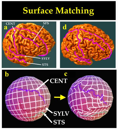
Probabilistic Atlases of Cortical Patterns. The random vector field approach is a general strategy to construct population-based atlases of the brain (Thompson and Toga, 1997). Briefly, given a 3D MR image of a new subject, a high-resolution parametric surface representation of the cerebral cortex is automatically extracted (Fig. 3). The algorithm then calculates a set of high-dimensional volumetric maps, elastically deforming this surface into structural correspondence with other cortical surfaces, selected one by one from an anatomic image database. The family of volumetric warps so constructed encodes statistical properties of local anatomical variation across the cortical surface. Specialized strategies elastically deform the sulcal patterns of different subjects into structural correspondence, in a way which matches large networks of gyral and sulcal landmarks with their counterparts in the target brain (Fig. 3). Differences in the serial organization of cortical gyri prevent exact gyrus-by-gyrus matching of one cortex with another, but an important intermediate goal has been to match a comprehensive network of sulcal and gyral elements which have consistent topology across subjects (Drury et al., 1996; Thompson et al., 1997). A probability space of random transformations, based on the theory of anisotropic Gaussian random fields, is then used to encode information on complex variations in gyral and sulcal topography from one individual to another (Fig. 5). Confidence limits in stereotaxic space are determined, for cortical surface points in the new subject's brain, enabling the creation of color-coded probability maps to highlight and quantify regional patterns of deformity in the anatomy of new subjects.
Pathology Detection. In view of the clear clinical and research applications, mathematical methods have been developed to create probabilistic measures of anatomic variation which are capable of detecting pathology. We (Thompson et al., 1996b) proposed a simple type of anomaly detection framework. Deformation fields are calculated which match elements of the deep surface anatomy in a range of N normal subjects. After affine components of the deformation fields are factored out, deviations from the mean deformation field are modeled, for small N, as a Hotelling's T-squared distributed random field, or for very large N, as a chi-squared distributed random field with 3 degrees of freedom, defined at nodes (u,v) in parametric mesh models of the anatomy of new subjects. Thompson et al. (1997) and Cao and Worsley (1998) proposed the use of a Hotelling's T-squared random field to detect abnormal deformations between groups, modeling the 3D deformation field, at each location, by a trivariate normal distribution with an arbitrary covariance tensor that allows for correlation between the coordinates. A T-squared or F statistic which indicates evidence of significant difference in deformations between the groups is calculated at each lattice location in a 3D image or parameterized 3D surface, to form a statistic image. Under the null hypothesis of no abnormal deformations, the statistic image is approximated by a T-squared random field. The global maximum of the random field can be used to test the hypothesis of no change (Worsley, 1994a,b; Cao and Worsley, 1998). Random field approaches, some of which are now widely used in packages for analyzing functional brain images (Friston et al., 1995), use the Euler characteristic (EC) of the excursion sets of a random field as an estimator of the number of local non-zero signal components, above a given threshold in a statistic image. They also use the expected value of the EC as an approximate p-value for the local maximum (Worsley, 1994a,b). Probabilistic atlases based on random deformation fields have been used to assess gender-specific differences in the brain (Cao and Worsley, 1998), and to detect structural abnormalities in neurodegenerative disorders such as Alzheimer's disease (Thompson et al., 1997).
Shape Theory Approaches. Deformation fields expressing neuroanatomic differences have also been analyzed using Procrustes methods, developed for the statistical analysis of biological shape (Bookstein, 1989; 1997). In Procrustes methods, affine components of neuroanatomic difference are factored out not by stereotaxic alignment, but by rotating and scaling configurations of point landmarks in each subject into least-squares correspondence with a Procrustes mean shape. Residual deformations which reflect individual change or anatomic difference are then expressed in terms of an orthogonal system of principal deformations derived from the bending energy matrix of the operator which governs the deformation (Bookstein, 1997). Of particular relevance are methods used to define a mean shape in such a way that departures from this mean shape can be treated as a linear process. Linearization of the pathology detection problem, by constructing Riemannian shape manifolds and their associated tangent spaces, allows the use of conventional statistical procedures and linear decomposition of departures from the mean to characterize shape change. These approaches have been applied to detect structural anomalies in schizophrenia (DeQuardo et al., 1996; Bookstein, 1997).
Pattern-Theoretic Approaches. In a related approach based on pattern theory (Grenander and Miller, 1994), a spectral approach to representing anatomic variation is developed. This approach, like the approaches described above, builds on the framework of deformable atlases by representing variation in terms of probabilistic transformations applied to deformable neuroanatomic templates. Deformation maps expressing variations in normal anatomies are calculated, with a non-linear registration procedure based on continuum-mechanics. In this formulation, the deformational behavior of each subject's anatomy, driven into correspondence with other anatomies, is expressed as a system of partial differential equations. The equations are governed by a differential operator controlling the way in which one anatomy is deformed into the other, and its properties can be used to make the deformation reflect the mechanical properties of deformable elastic or fluid media. Common choices of the differential operator are the Laplacian (Joshi et al., 1995), biharmonic (Bookstein, 1989) and Cauchy-Navier operator (Bajcsy and Kovacic, 1989; Gee et al., 1993, 1995; Miller et al., 1993; Christensen et al., 1996; Davatzikos, 1996; Thompson and Toga, 1998). Each deformation map is then expanded in terms of the eigenfunctions of the governing operator, and Gaussian probability measures are defined on the resulting sequences of expansion coefficients. Currently being tested as a framework for representing anatomic variation, this pattern-theoretic approach builds on the framework of deformable atlases and shows promise in the automated detection of pathology.
Paul Thompson
| RESUME| E-MAIL ME| PERSONAL HOMEPAGE| PROJECTS |
|---|