Received Dec. 18, 1995; revised March 19, 1996; accepted April 5, 1996.
Laboratory of Neuro Imaging, Department of Neurology, Division of Brain Mapping, UCLA School of Medicine, Los Angeles, California 90095
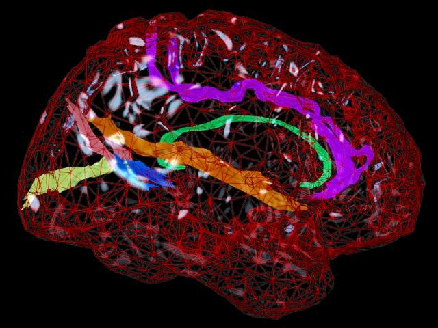
Morphometric variance of the human brain is qualitatively observable in surface features of the cortex. Statistical analysis of sulcal geometry will facilitate multisubject atlasing, neurosurgical studies, and multimodality brain mapping applications. This investigation describes the variability in location and geometry of five sulci surveyed in each hemisphere of six postmortem human brains placed within the Talairach stereotaxic grid. The sulci were modeled as complex internal surfaces in the brain. Heterogeneous profiles of three-dimensional (3D) variation were quantified locally within individual sulci.
Whole human heads, sectioned at 50 µm, were digitally photographed and high-resolution 3D data volumes were reconstructed. The parieto-occipital sulcus, the anterior and posterior rami of the calcarine sulcus, the cingulate and marginal sulci, and the supracallosal sulcus were delineated manually on sagittally resampled sections. Sulcal outlines were reparameterized for surface comparisons. Statistics of 3D variation for arbitrary points on each surface were calculated locally from the standardized individual data. Additional measures of surface area, extent in three dimensions, surface curvature, and fractal dimension were used to characterize variations in sulcal geometry.
Paralimbic sulci exhibited a greater degree of anterior-posterior variability than vertical variability. Occipital sulci displayed the reverse trend. Both trends were consistent with developmental growth patterns. Points on the occipital sulci displayed a profile of variability highly correlated with their 3D distance from the posterior commissure. Surface curvature was greater for the arched paralimbic sulci than for those bounding occipital gyri in each hemisphere. On the other hand, fractal dimension measures were remarkably similar for all sulci examined, and no significant hemispheric asymmetries were found for any of the selected spatial and geometric parameters. Implications of cortical morphometric variability for multisubject comparisons and brain mapping applications are discussed.
Key words: brain mapping; cortex; stereotaxic methods; sulcus; 3D image reconstruction; morphometry
Paul Thompson
| RESUME| E-MAIL ME| PERSONAL HOMEPAGE| PROJECTS |
|---|