Paul M. Thompson, David MacDonald, Michael S. Mega, Colin J. Holmes, Alan C. Evans, Arthur W. Toga
Laboratory of Neuro Imaging and Alzheimer's Disease Center, Department of Neurology, Division of Brain Mapping, UCLA School of Medicine, Los Angeles, California 90095
and
Montreal Neurological Institute, McGill University, Quebec, Canada
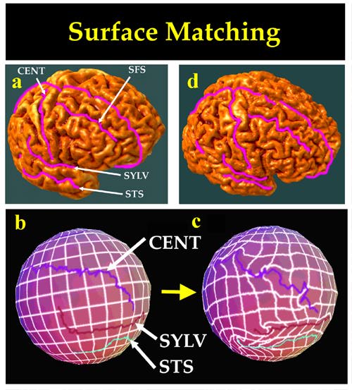
Method. Given a 3D MR image of a new subject, a high-resolution surface representation of the cerebral cortex is automatically extracted. The algorithm then calculates a set of high-dimensional volumetric maps, fluidly deforming this surface into structural correspondence with other cortical surfaces, selected one by one from an anatomic image database. The family of volumetric warps so constructed encodes statistical properties of local anatomical variation across the cortical surface. Additional strategies are developed to fluidly deform the sulcal patterns of different subjects into structural correspondence. A probability space of random transformations, based on the theory of anisotropic Gaussian random fields, is then used to encode information on complex variations in gyral and sulcal topography from one individual to another. A complete system of 256x256 probability density functions is computed to reflect the observed variability in stereotaxic space of the points whose correspondences are found by the warping algorithm. Confidence limits in stereotaxic space are determined for cortical surface points in the new subject's brain.
Results. Color-coded probability maps are generated, which highlight and quantify regional patterns of deformity in the anatomy of new subjects. These maps indicate locally the probability of each anatomic point being as unusually situated, given the distributions of corresponding points int he scans of normal subjects. 3D MRI volumes are analyzed, from subjects with clinically-determined Alzheimer's Disease and age-matched normal subjects.
Conclusion. Applications of the random fluid-based probabilistic atlas include the transfer of multi-subject 3D functional, vascular, and histologic maps onto a single anatomic template, the mapping of 3D atlases onto the scans of new subjects, and the rapid detection, quantification, and mapping of local shape changes in 3D medical images in disease and during normal or abnormal growth and development.
 Detection of Abnormal Brain Structure: |
 Same Map Constructed for an Age-Matched |
Index Terms. Brain, Mapping-Image Registration-Atlas and Atlases-Stereotaxic Imaging-Magnetic resonance imaging-Image reconstruction, three-dimensional-Morphometry.
Paul Thompson
| RESUME| E-MAIL ME| PERSONAL HOMEPAGE| PROJECTS |
|---|