Michael S. Mega, Sylvia S. Chen, Paul M. Thompson, Roger P. Woods, Timur J. Karaca, Abhishek Tiwari, Harry V. Vinters, Gary W. Small, Arthur W. Toga
Laboratory of Neuro Imaging, Department of Neurology, Division of Brain Mapping, UCLA School of Medicine, Los Angeles, California 90095
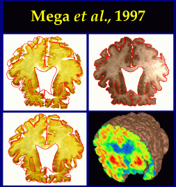
The association between [18F]fluorodeoxyglucose positron emission tomography (FDG-PET) counts obtained 8 hours before death and neurofibrillary tangle (NFT) staining density in a patient with Alzheimer's disease (AD) was evaluated. In our patient FDG-PET counts were globally decreased with a greater focal deficit in the left medial temporal region independent of volume loss. After death, whole-brain sections derived from cryomacrotome sectioning were stained for NFTs by the Gallyas method, and elastically warped into their native space enabling registration with premortem FDG-PET data. Gallyas staining density was localized in the paralimbic cortex of the basal forebrain, medial temporal, and orbital frontal regions. The poor correlation between NFT staining density and hypometabolism on FDG-PET implicates alternate mechanisms underlying the metabolic defect in AD.
Paul Thompson
| RESUME| E-MAIL ME| PERSONAL HOMEPAGE| PROJECTS |
|---|