3D Cortical Pattern Variability in
Schizophrenic Patients and Matched Normal Subjects.
These maps are made using new mathematical and computational
strategies for cortical mapping and for creating
population-based brain atlases.
Narr KL, Sharma T, Moussai J, Zoumalan CI, Cannestra AF, Thompson PM, Toga AW
Laboratory of Neuro Imaging, Dept. Neurology, Division of Brain Mapping,
UCLA School of Medicine, Los Angeles CA 90095, USA,
and
Institute of Psychiatry, London
3D Cortical Pattern Variability in
Schizophrenic Patients and Matched Normal Subjects.
These maps are made using new mathematical and computational
strategies for cortical mapping and for creating
population-based brain atlases.
Introduction. Neuroanatomic abnormalities are characteristic in schizophrenic populations. Cerebral anomalies reported include alterations in sulco-gyral patterns [1] and alterations in cerebral asymmetries [2],[3]. To map group variability, displacement and asymmetry of sulci and surrounding anatomy, 3D cortical extractions from MR scans were used and brought into register with the aid of surface warping algorithms that enable averaging of equivalent cortical regions across subjects [4].
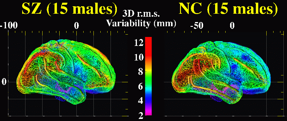
Methods. High-resolution (256x256x124; 1.5mm separation) T1-weighted MR images were acquired from schizophrenic patients and controls (N=15 per group; all males) matched for age, educational level, and handedness. Each 3D volume was aligned and scaled according to Talairach AC-PC distance (Talairach and Tournoux, 1988). Complex 3D shape representations of the cortex were then extracted [4] and delineated by manually tracing major fissures and sulci on highly magnified hemispheric surfaces following anatomic criteria [5]. Sulcal curves included: inferior and superior frontal and temporal sulci; central; postcentral; intraparietal; occipital-temporal; collateral; and olfactory sulci and Sylvian fissures in addition to midline curves separated by anatomic boundaries [7]. Variability and displacement of uniformly re-parameterized sulcal anatomy were mapped by creating average surface mesh models within each population [6]. Cortical surface variability was then calculated using average landmark curves as anchors to drive the 3D surface renderings from each subject into correspondence. Briefly, parametric meshes modeling cortical surfaces are translated from spherical tensor to planar maps retaining 3D position information encoded in color. A rectangular grid placed over each planar color map allows averaging of grid-points from the same cortical locations using a flow field. Finally, group averaged cortical models are translated back into 3D retaining information about individual cortical patterns. Patterns of variability in each group are visualized as the root mean square magnitude of displacement vectors from mapping each subject onto the average group mesh [7]. Alterations in structural asymmetries were measured in stereotaxic co-ordinates derived from the sulcal curves.
Results. Variability maps revealed different profiles of sulcal variability across groups and distinct asymmetries in the inferior and superior temporal sulci, Sylvian fissures and post central sulci within groups (see figure above). Variability was increased in superior extents of the temporal sulci including the Sylvian fissure. This was more extreme in right perisylvian cortices and in frontal and intraparietal sulci in patients. Overall, in both groups, maps of cortical surface variability indicated increased individual differences in neopallial association areas in respect to phylogenetically older areas of cortex. Talairach AC-PC scaling was examined as a possible covariate for measures of sulcal asymmetry but did not significantly contribute to the variance. Mixed 2 (Diagnosis) x 2 (Hemisphere) MANOVAs were thus used without corrections of AC-PC distance to examine differences in superior and posterior extents and curvature of temporal sulci and anterior extents and curvature of the postcentral sulci. Significant asymmetries were found in the posterior extents of the inferior and superior temporal sulci and Sylvian fissure, (F (1,28) = 14.04; 16.27 and 41.43 respectively, p < 0.001) with left hemisphere sulci extending more posteriorly. A significant asymmetry was also detected for anterior postcentral sulcal extents, F (1,28) = 13.42, p = 0.001 with anterior displacement on the right. There were no significant effects of Diagnosis.
Conclusions. Variability maps and statistical analyses revealed distinct asymmetries of sulcal geometry in both schizophrenic patients and nonpsychiatric controls. These results argue against patterns of altered asymmetries in temporal regions in schizophrenic patients. There were however different patterns of cortical surface variability across groups implicating regional abnormalities in schizophrenic patients.
References. [1] Kikinis et al., Neuroscience Letters. 182:7-12. 1994. [2] Bartley et al., Biol. Psychiatry, 34:853-63. 1993. [3] Lawrie & Abukmeil, Br. J. Psychiatry, 172:110-20.1998. [4] Thompson et al., J. Comp. Assist. Tomography, 21:567-81. 1997. [5] Ono et al., Atlas of the Cerebral Sulci. Stuttgart: Theime. 1990. [6] Thompson et al., J. Neuroscience, 16:4261-74. 1996. [7] Thompson et al., 1999, Human Brain Mapping 8(4), Sept. 1999.
Grant Support: (to P.T. and A.W.T.): NIMH/NIDA (P20 MH/DA52176), P41 NCRR (RR13642); (A.W.T.): NLM (LM/MH05639), NSF (BIR 93-22434), NCRR (RR05956) and NINCDS/NIMH (NS38753).
Paul Thompson
| RESUME| E-MAIL ME| PERSONAL HOMEPAGE| PROJECTS |
|---|