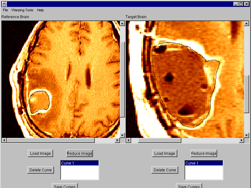
Axial MRI Scan of a Patient with Glioblastoma Multiforme.
These scans can also be
tissue-classified
by a
computer vision algorithm
to identify specific tissue types.

Tumor Growth in a Patient with Glioblastoma Multiforme.
Wong GK, Haney S, Thompson PM, Alger JR, Cloughesy TF, Toga AW
Laboratory of Neuro Imaging, Dept. Neurology, Division of Brain Mapping,
UCLA School of Medicine, Los Angeles CA 90095, USA,
UCLA Dept. of Radiological Sciences, and
Neuro-Oncology Program, UCLA School of Medicine

Axial MRI Scan of a Patient with Glioblastoma Multiforme.
These scans can also be
tissue-classified
by a
computer vision algorithm
to identify specific tissue types.

Tumor Growth in a Patient with Glioblastoma Multiforme.
Summary. As part of a longitudinal evaluation of tumor growth in patients with glioblastoma multiforme (GBM), a battery of pattern recognition and 3-dimensional image analysis algorithms were applied to serial MRI scans. We evaluated the ability of semi-automated tissue classification algorithms
(1) to quantify 3D volumes and accurately delineate the boundaries of the tumor, necrotic, and gadolinium-enhancing tissue as well as peritumoral edema, adjacent white matter, and cystic compartments; and
(2) to detect and map changes in compartments over time.
Methods. 3D T1-weighted (256x256 resolution; 3 mm spacing) SPGR MRI volumes were acquired before and after the administration of gadolinium contrast from 11 patients with GBM. Patients ranged in age from 4 to 54 at first scan. Scans acquired over a period up to 3.5 years, at intervals ranging from 6 weeks to 6 months, were digitally converted into Talairach stereotaxic space using 6-parameter rigid transformations. 160 tags (170 when a cystic component was present) per anatomical region were then used to determine a Gaussian mixture distribution reflecting the intensities of specific tissue classes at each time-point in the scan series. A nearest neighbor algorithm was used to differentiate tissue types. Tissue maps were manually adjusted to better delineate class boundaries and the results of automated and manual segmentations were compared. Volumetric changes in time ranged from -73% to +2,600 % corresponding to a halving time of 130 days and a doubling time of 13.8 days.
Conclusions. These preliminary data suggest that single scan tissue classification shows promise in defining tissue types. Multispectral classifcation, however, may be required to define peritumoral edema. Tissue maps, in conjunction with other growth rate data, will provide a valuable adjunct to clinical, spectroscopic, and histopathologic indices in mapping rates of change for different tumor attributes. They may also be useful in evaluating therapeutic response in individual patients and clinically-defined groups.
Grant Support: (to P.T. and A.W.T.): NIMH/NIDA (P20 MH/DA52176), P41 NCRR (RR13642); (A.W.T.): NLM (LM/MH05639), NSF (BIR 93-22434), NCRR (RR05956) and NINCDS/NIMH (NS38753).
Paul Thompson
| RESUME| E-MAIL ME| PERSONAL HOMEPAGE| PROJECTS |
|---|