|
|
(Click here to Return to the Abstract of the Paper)
(Click here to Return to the List of Current Projects)
Probabilistic Brain Atlases:
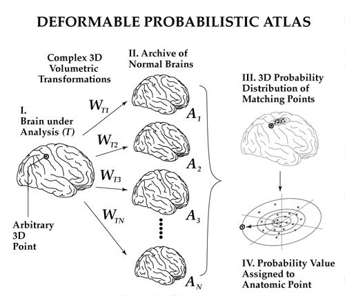
(1) Strategy for Creating a Population-Based Brain Atlas.
A family of high-dimensional volumetric warps relating a new subject's
3D MRI scan to each normal scan in a
brain image database is calculated (I-II,
above) and then used to quantify local structural variations.
Differences
in cortical, ventricular and deep sulcal topography are recorded in the
form of vector
field transformations in 3D stereotaxic space which drive
both subcortical anatomy and the gyral/sulcal
patterns of different subjects
into register. The resulting family of warps encodes the
distribution in
stereotaxic space of anatomic points that correspond across a normal population
(III),
and their
dispersion is used to determine the likelihood (IV) of
local regions of the new subject's anatomy being
in their actual configuration.
Easily interpretable, color-coded topographic maps are generated to
highlight
regional patterns of deformity in the anatomy of the new subject. Abnormal structural patterns
are quantified locally,
and mapped in three dimensions. |
|
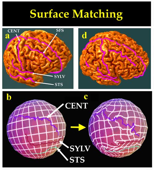
(2) Scheme for Matching Cortical Regions with
High-Dimensional Transformations and Color-Coded Spherical Maps. High-resolution
surface models of the
cerebral cortex were extracted in parametric form,
using the active surface algorithm of
(MacDonald et al., 1993, 1994).
This means that a continuous, invertible one-to-one mapping is always
available
between points on the cortical surface, (a), and their counterparts on
the surface of a sphere.
No 3D information is lost in this data representation
scheme, as each point in the spherical map, (b), is
color-coded with a
color value which accurately and uniquely represents the location of its
counterpart
on the convoluted surface model (a) in 3D stereotaxic space.
To preserve accuracy, floating point triplets,
representing cortical surface
point locations in stereotaxic space, are color-coded at 16 bits per channel
to form an image of the parameter space in RGB color image format. To find good matches between cortical
regions in different subjects [(a)/(d)],
we first derive a colorized spherical map for each respective surface
model
(b and c) and perform the matching process in the angular parametric space.
When spherical maps are made
from two different cortical surfaces, the
respective sulci will be in different positions in each spherical
map (b
and c), reflecting their different locations on the folded brain surface
[shown here in pink,
a and d]. Using a complex vector-valued flow
field defined on the
sphere (c), the system of sulcal curves in one spherical map can be driven
into exact
correspondence with their counterparts in the target spherical
map, guiding the transformation of the adjacent
regions. A spatially accurate,
anatomically driven warping algorithm
(Thompson and Toga, 1996),
calculates the high-dimensional deformation field (typically with
65536x3 ~
200,000 degrees of freedom) which reconfigures
the starting spherical map, and the networks of curves
embedded within
it, into the shape of their counterparts in the target spherical map. This
transformation is illustrated in (c) by its effect on a uniform
grid, ruled over
the starting spherical map and passively carried along
in the resultant deformation. Notice
the complex reconfiguration of sulcal
landmarks, and how they drive the deformation of the
surrounding cortex,
allowing for complex profiles of dilation and contraction of the surface
into the shape of the target surface. Note the complex non-linear flow in superior temporal
regions, as the superior temporal sulcus (STS) extends
further posteriorly in the target brain,
and the posterior upswing of the
Sylvian fissure (SYLV) is more pronounced in the reference
brain (a) than
in the target (d). Outlines are also shown for the superior frontal sulcus
(SFS), and for the central sulcus (CENT) which is less convoluted in
the reference brain
than in the target. Because the color-coded spherical
maps index cortical surface locations
in 3-D, the transformation of one
spherical map to another can be recovered in 3D stereotaxic
space as a
displacement of points in one subject's cortex onto their counterparts
in the cortex
of another subject. Matching can therefore be driven by a network of anatomically significant
surface features. High spatial accuracy
of the match is guaranteed in regions of particular
functional significance
or structural complexity, such as sulcal curves, lobar and
cytoarchitectural
boundaries, and critical functional landmarks.
|
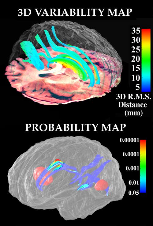 |
(3) Distortions in Brain Architecture Induced by
Tumor Tissue:
Probability Maps for Major Sulci in Both Hemispheres. (Top)
3D r.m.s. variability maps are shown for major
occipital and paralimbic
sulci; (bottom) color-coded probability maps quantify the impact of two
focal
metastatic tumors (illustrated in red) on the supracallosal, parieto-occipital,
and anterior and posterior
calcarine sulci in both hemispheres. |