Paul M. Thompson, Katherine L. Narr, Rebecca E. Blanton, Arthur W. Toga
Laboratory of Neuro Imaging, Department of Neurology, Division of Brain Mapping, UCLA School of Medicine, Los Angeles, California 90095

The rapid growth in brain imaging technologies has been matched by an extraordinary increase in the number of investigations focusing on analyzing callosal structure and function. Surgical transection of the callosum in humans provides evidence that it functions to communicate perceptual, cognitive, mnemonic, learned and volitional information between the two brain hemispheres (Bogen et al., 1965). Given the importance of sensory, motor and cognitive callosal relay between hemispheres, it is not surprising that this anatomic region has been a focus of studies examining structural and functional neuropathology.
Disease. In disease, effects on callosal structure are observable at both cellular and gross anatomic scales (Innocenti, 1994). We and other groups (Thompson et al., 1998; Hofmann et al., 1995; Vermersch et al., 1996; Janowsky et al., 1996) have investigated callosal abnormalities in Alzheimer's Disease, and callosal anomalies have also been reported in multi-infarct dementia (Yoshii et al., 1990), schizophrenia (Woodruff et al., 1995; DeQuardo et al., 1996), attention deficit hyperactivity disorder (Giedd et al., 1994; Baumgardner et al., 1996), relapsing-remitting multiple sclerosis (Pozzilli et al., 1991), and a range of neurodevelopmental disorders and dysplasias (Sobire et al., 1995).
Intense Controversy over Reports of Structural Differences. Analysis of regional neuroanatomy and callosal structure are key factors in the radiologic assessment of a wide range of neurological disorders. Nonetheless, extreme variations in brain structure make it difficult to design computerized strategies that detect and classify abnormal structural patterns. Intense controversy exists on the question of whether different callosal regions undergo selective changes in each of these disease processes. Additional controversy surrounds studies which have identified specific differences in callosal structure related to sex (DeLacoste-Utamsing and Holloway, 1982), handedness (Witelson, 1985), IQ (Strauss et al., 1994), musical ability (Schlaug et al., 1995), and in studies comparing monozygotic and dizygotic twins (Oppenheim et al., 1989). Anatomically-specific relationships between callosal structure, cortical asymmetry, and a range of cognitive measures have generated great interest, in view of the light they shed on the organization of brain function and the nature of interhemispheric communication.
Summary of Our Chapter. In our review of current neuroimaging research on callosal structure, we emphasize the many recent mathematical and methodological advances in the field. Imaging and computational methods are discussed for analyzing and understanding the complex dynamic processes that affect callosal anatomy in the healthy and diseased brain.
Growth Patterns. In many ways, static representations of callosal structure are ill-suited to determining the dynamic effects of development and disease. The callosum undergoes profound changes in morphology, fiber composition and myelination during brain development, and these patterns are dramatically altered in disease. Later in the chapter, we discuss methods for mapping dynamic patterns of growth at the corpus callosum. Several algorithms are used to create 4-dimensional quantitative maps of complex growth patterns across the corpus callosum, based on time-series of pediatric MRI scans (Thompson et al., 1998; Thompson and Toga, 1998). Mathematical techniques are designed to analyze local growth rates, revealing the local magnitude and principal directions of dilation or contraction. Serial scanning of human subjects, coupled with computational methods for analyzing the changing callosal geometry, show promise in enabling disease and growth processes to be tracked in their full spatial and temporal complexity.
Probabilistic Brain Atlases. We also review progress in constructing a probabilistic reference system for the human brain, which can be used to analyze callosal anatomy (Mazziotta et al., 1995; Thompson et al., 1997, 1998). MRI-based image archives from large human populations are stratified into subpopulations according to age, disease state, gender and other demographic criteria, to produce population-specific representations of anatomy. The resulting brain atlases encode information on population variability, with the goal of identifying group-specific patterns of brain structure. Computerized strategies are developed to (1) detect abnormal callosal structure in disease, (2) relate detected callosal anomalies to changes in the 3-dimensional patterns of cortical asymmetry and structural variability, and (3) relate callosal changes found in development and aging to changes found in Alzheimer's Disease and schizophrenia.
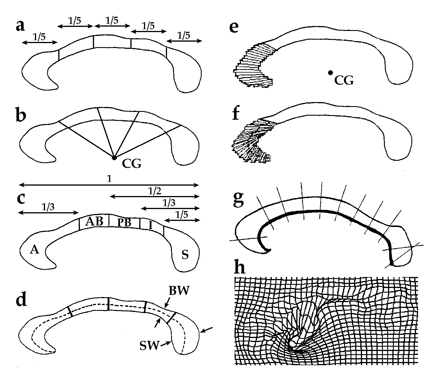
Acknowledgment. The figure (above) shows common partitioning schemes for regional analysis of the corpus callosum, and is adapted from diagrams created by Witelson (1989); Clarke et al., (1989); Clarke and Zaidel, 1994; Rajapakse et al., 1996; and Seivenart et al., 1997.
To obtain a reprint of our chapter, please send me an e-mail, and I'll be happy to send you a copy!
Key Words: Brain Mapping, 3D, corpus callosum, Magnetic Resonance Imaging
Paul Thompson
| RESUME| E-MAIL ME| PERSONAL HOMEPAGE| PROJECTS |
|---|