Paul Thompson's Research Publications
Growth Rates, MR Spectroscopy: Prognostic Markers for Glioblastoma Multiforme
Society for Neuro-Oncology, Chicago, Nov. 2000
Haney S, Thompson PM, Cloughesy
TF, Alger JR, Frew AJ, Toga AW
Laboratory of Neuro Imaging, Department of Neurology, Division of
Brain Mapping;
Neuro-Oncology Program, The Henry E. Singleton Brain Cancer Research Program;
Dept. of Radiological Sciences; UCLA School of Medicine, Los Angeles,
CA 90095
|
ABSTRACT
|
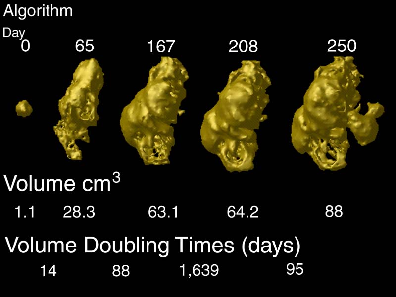
Mapping Rates of Tumor Growth. Rates of tumor growth in GBM patients
can be tracked using a combination of image analysis algorithms. Maps of contrast-enhancing
tumor can be generated using tissue classifiers. Growth rates can be calculated from these
maps, and monitored during therapy. Here growth rates are expressed in terms of equivalent
doubling times, which indicate how long it would take for the tumor to double in volume,
based on the changes observed in an interval between MRI scans.
Introduction
As part of an ongoing study of patients with malignant gliomas, we sought
to determine whether
imaging markers such as contrast enhancing tissue, edema and necrosis,
MR spectroscopic
measures, and other factors were prognostically significant.
Prognostic markers offer
valuable information for the physician providing treatment, for the patient
himself, especially in
terms of making decisions regarding quality of life issues, and for the
researcher designing clinical
studies targeting specific subgroups of patients. A variety of potential
prognostic information,
such as tumor location, presenting symptoms, and time to recurrence were
also tracked as part
of this study. Imaging measures may function as dynamic
markers, providing prognostic data
which may be revised during the course of therapy.
Methods
16 patients
with histopathologically
confirmed diagnosis of glioblastoma multiforme (GBM), and with tumors that
were neither midlinear
or infratentorial, were serially scanned. Measures of edema, necrosis
and contrast-enhancing
tissue were derived from T2-weighted and gadolinium enhanced T1-weighted
SPGR (spoiled
Grass; 256x256, 3mm spacing, no gap) MRIs. MR spectroscopy measures
were also acquired.
Volumetric analysis was performed using a 3D tissue segmentation algorithm
to guide manual
segmentation. Volumetric data was then compared across time.
Growth rates for contrast
enhancing tissue and CHO/CRE (choline/creatine) measures were examined.
Results
Preliminarily, multivariate analysis indicated a significant
negative relationship
between growth of contrast enhancing tumor tissue and survival (p<0.03).
Values of CHO/CRE > 1.8 were
significantly negatively related to survival. Age, a well-known
prognostic marker, was also
significantly negatively related to survival (p<0.01). Analysis
of other factors is ongoing.
Conclusion
Multivariate analysis indicates that growth rates of contrast-enhancing tissue,
CHO/CRE measures and age are prognostically significant. These prognostic factors, as
well as others, are valuable for the
patient and physician in making decisions regarding therapy choice,
and for reserchers designing
clinical trials.
Related Publications
(back
to main list)
Contact Information
Mail:
Paul Thompson, Ph.D.
Assistant Professor of Neurology
Lab of Neuro Imaging and Brain Mapping Division
4238 Reed Neurology
710 Westwood Plaza
UCLA Medical Center
Los Angeles CA 90095-1761, USA.
E-mail:
thompson@loni.ucla.edu
Tel: (310)206-2101
Fax: (310)206-5518