Proc. IEEE International Conference on Computer
Vision and Pattern Recognition, and
Workshop on Mathematical Methods in
Biomedical Image Analysis, Hilton Head Island, South Carolina, June 11-12
2000, pp. 227-234.
Paul M. Thompson, Michael S. Mega, and Arthur W. Toga
Laboratory of Neuro Imaging, Dept. Neurology, Division of Brain Mapping, and
Alzheimer's Disease Center, UCLA School of Medicine, Los Angeles CA 90095, USA
E-mail:
thompson@loni.ucla.edu
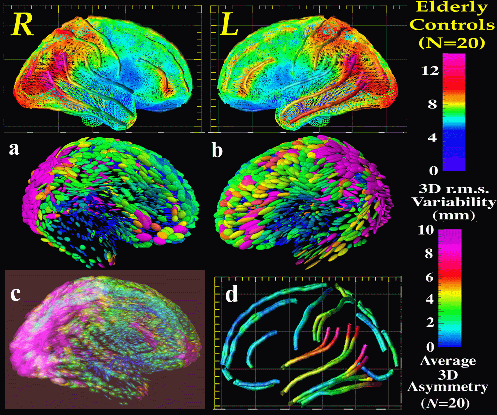
Atlases of the human brain, in health and disease, provide a comprehensive framework for understanding brain structure and function. The complexity and variability of brain structure, especially in the gyral patterns of the human cortex, present challenges in creating standardized brain atlases that reflect the anatomy of a population. This paper introduces the concept of a population-based, disease-specific brain atlas that can reflect the unique anatomy and physiology of a particular clinical subpopulation. Based on well-characterized patient groups, disease-specific atlases contain thousands of structure models, composite maps, average templates, and visualizations of structural variability, asymmetry and group-specific differences. They correlate the structural, metabolic, molecular and histologic hallmarks of the disease. Rather than simply fusing information from multiple subjects and sources, new mathematical strategies are introduced to resolve group-specific features not apparent in individual scans. High-dimensional elastic mappings, based on covariant partial differential equations, are developed to encode patterns of cortical variation. In the resulting brain atlas, disease-specific features and regional asymmetries emerge that are not apparent in individual anatomies. The resulting probabilistic atlas can identify patterns of altered structure and function, and can guide algorithms for knowledge-based image analysis, automated image labeling, tissue classification, data mining and functional image analysis.
Paul Thompson, Ph.D.
| RESUME| E-MAIL ME| PERSONAL HOMEPAGE| PROJECTS |
|---|