Book Chapter in: Isaac Bankman, Raj Rangayyan, Alan Evans, Roger Woods, Elliot Fishman, Henry Huang
[eds.],
Handbook of Medical Image Processing, Academic
Press, 1999 [in press]
Paul M. Thompson and Arthur W. Toga
Laboratory of Neuro Imaging, Department of Neurology, Division of Brain Mapping, UCLA School of Medicine, Los Angeles, California 90095

Image registration is central to many of the challenges in brain imaging today. Initially developed as an image processing subspecialty to geometrically transform one image to match another, registration now has a vast range of applications.
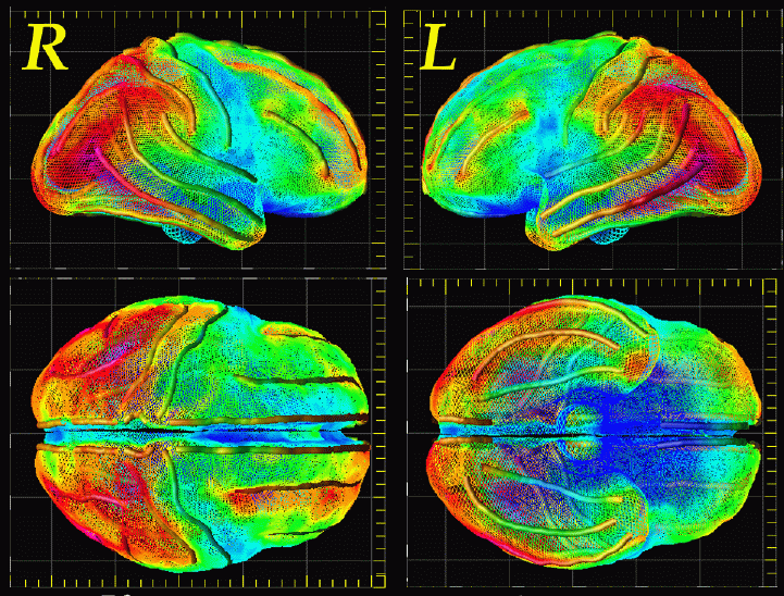
In this chapter, we review the registration strategies currently used in medical imaging, with a particular focus on their ability to detect and measure differences. These include methods developed for automated image labeling and for pathology detection in individuals or groups. We show that these algorithms can serve as powerful tools to investigate how regional anatomy is altered in disease, and with age, gender, handedness and other clinical or genetic factors. Registration algorithms can encode patterns of anatomic variability in large human populations, and can use this information to create disease-specific, population-based brain atlases. They may also fuse information from multiple imaging devices to correlate different measures of brain structure and function. Finally, registration algorithms can even measure dynamic patterns of structural change during brain development, tumor growth, or degenerative disease processes.
Pattern recognition algorithms for automated identification of brain structures can benefit greatly from encoded information on anatomic variability. Registration algorithms, applied in a probabilistic framework, also offer a new method to examine abnormal brain structure. Probability maps can be combined with anatomically-driven elastic transformations that associate homologous brain regions in an anatomic database. This provides the ability to perform morphometric comparisons and correlations in 3D between a given subject's MR scan and a population database, or between population sub-groups stratified by clinical or demographic criteria.
To obtain a reprint of our chapter, please send me an e-mail, and I'll be happy to send you a copy!
Key Words: Brain Mapping, 3D, image registration, deformable templates, elastic matching, Magnetic Resonance Imaging
Paul Thompson
| RESUME| E-MAIL ME| PERSONAL HOMEPAGE| PROJECTS |
|---|