Paul M. Thompson, Michael S. Mega, and Arthur W. Toga
Laboratory of Neuro Imaging, and Alzheimer's Disease Center, Department of Neurology, Division of Brain Mapping, UCLA School of Medicine, Los Angeles, California 90095
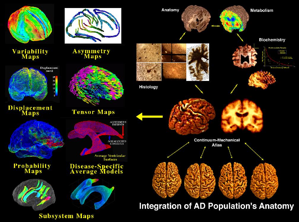
This chapter introduces the topic of a disease-specific brain atlas (see image above). This type of atlas is designed to reflect the unique anatomy and physiology of a particular clinical subpopulation (Thompson et al., 1997, 1998, 1999; Mega et al., 1997, 1998, 1999; Narr et al., 1999). Based on well-characterized patient groups, these atlases contain thousands of structure models, as well as composite maps, average templates, and visualizations of structural variability, asymmetry and group-specific differences. They act as a quantitative framework that correlates the structural, metabolic, molecular and histologic hallmarks of the disease (Mega et al., 1997). Additional algorithms are described that use information stored in the atlas to recognize anomalies and label structures in new patients. Because they retain information on group anatomical variability, disease-specific atlases are a type of probabilistic atlas specialized to represent a particular clinical group. The resulting atlases can identify patterns of altered structure or function, and can guide algorithms for knowledge-based image analysis, automated image labeling, tissue classification, and functional image analysis.
We present data from several on-going projects, whose goal is to create disease-specific atlases of the brain in Alzheimer's Disease, schizophrenia, and several neurodevelopmental disorders. Since current brain templates poorly represent the anatomy of these clinical populations, the resulting atlases offer a framework to investigate each disease. Pathological change can be tracked over time, and disease-specific features resolved. Rather than simply fusing information from multiple subjects and sources, new mathematical strategies are introduced to resolve group-specific features not apparent in individual scans.
To obtain a reprint of our chapter, please send me an e-mail, and I'll be happy to send you a copy!
Key Words: Brain Mapping, 3D, Alzheimer's Disease, schizophrenia, brain development, brain asymmetry, cortical organization, brain structure and function, Magnetic Resonance Imaging
Paul Thompson
| RESUME| E-MAIL ME| PERSONAL HOMEPAGE| PROJECTS |
|---|