
Cerebral Cortex 11(1):1-16, Jan. 2001
Paul M. Thompson, Michael S. Mega, Roger P. Woods, Chris I. Zoumalan, Chris J. Lindshield, Rebecca E. Blanton, Jacob Moussai, Colin J. Holmes, Jeffrey L. Cummings, and Arthur W. Toga
Laboratory of Neuro Imaging, Dept. Neurology, Division of Brain Mapping,
UCLA School of Medicine, Los Angeles CA 90095, USA,
and
UCLA Alzheimer's Disease Center


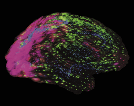
Cover Picture: Tensor maps reveal regional patterns in cortical variability. Striking differences are found, even among normal human subjects, in the gyral patterns of the cerebral cortex. Tensor maps (cover image) can reveal the preferred directions and the magnitude of anatomical variation, for different cortical regions. Color distinguishes regions of high variability (pink colors, in temporo-parietal cortices) from areas of low variability (blue colors, in olfactory gyri). Ellipsoidal glyphs are most elongated along directions in which variability is greatest across subjects. These nested ellipsoids, shown with varying transparency, represent statistical confidence regions in which cortical anatomy is found in a certain percentage of the population. The resulting information can be used to distinguish normal from abnormal cortical anatomy. See Thompson et al., pp. 1-16.
Summary. We report the first detailed population-based maps of cortical gray matter loss in Alzheimer's disease (AD), revealing prominent features of early structural change. New computational approaches were used to: (i) distinguish variations in gray matter distribution from variations in gyral patterns; (ii) encode these variations in a brain atlas (n = 46); (iii) create detailed maps localizing gray matter differences across groups. High resolution 3D magnetic resonance imaging (MRI) volumes were acquired from 26 subjects with mild to moderate AD (age 75.8 ± 1.7 years, MMSE score 20.0 ± 0.9) and 20 normal elderly controls (72.4 ± 1.3 years) matched for age, sex, handedness and educational level. Image data were aligned into a standardized coordinate space specifically developed for an elderly population. Eighty-four anatomical models per brain, based on parametric surface meshes, were created for all 46 subjects. Structures modeled included: cortical surfaces, all major superficial and deep cortical sulci, callosal and hippocampal surfaces, 14 ventricular regions and 36 gyral boundaries. An elastic warping approach, driven by anatomical features, was then used to measure gyral pattern variations. Measures of gray matter distribution were made in corresponding regions of cortex across all 46 subjects. Statistical variations in cortical patterning, asymmetry, gray matter distribution and average gray matter loss were then encoded locally across the cortex. Maps of group differences were generated. Average maps revealed complex profiles of gray matter loss in disease. Greatest deficits (20–30% loss, P < 0.001–0.0001) were mapped in the temporo-parietal cortices. The sensorimotor and occipital cortices were comparatively spared (0–5% loss, P > 0.05). Gray matter loss was greater in the left hemisphere, with different patterns in the heteromodal and idiotypic cortex. Gyral pattern variability also differed in cortical regions appearing at different embryonic phases. 3D mapping revealed profiles of structural deficits consistent with the cognitive, metabolic and histological changes in early AD. These deficits can therefore be (i) charted in a living population and (ii) compared across individuals and groups, facilitating longitudinal, genetic and interventional studies of dementia.
Paul Thompson, Ph.D.
| RESUME| E-MAIL ME| PERSONAL HOMEPAGE| PROJECTS |
|---|