Paul M. Thompson, Michael S. Mega, Rebecca E. Blanton, Jacob Moussai, Shahin Zohoori, Amir Goldkorn, Colin J. Holmes, David MacDonald, Gary W. Small, Jeffrey L. Cummings and Arthur W. Toga
Laboratory of Neuro Imaging and Alzheimer's Disease Center, Department of Neurology, Division of Brain Mapping, UCLA School of Medicine, Los Angeles, California 90095
and
Montreal Neurological Institute, McGill University, Montreal, Canada
Methods: High-resolution probabilistic atlases of the brain [1] in normal aging and AD were constructed from a reference archive of T1-weighted 256x256x170 resolution 3D MRI scans of 10 AD patients (age:71.9+/-10.9 yrs.) and 10 controls matched for age (72.9+/-5.6 yrs.), gender, educational level and handedness. Scans were digitally transformed into Talairach stereotaxic space. Connected systems of parametric surface meshes were used to model 33 structures including parieto-occipital, calcarine, cingulate, marginal, callosal sulci, Sylvian fissures, 14 ventricular regions, superior and inferior hippocampal surfaces and other major lobar and cytoarchitectural boundaries in 3 dimensions. High-dimensional volumetric maps [2] (with 0.1 billion degrees of freedom) were computed, fluidly reconfiguring the anatomy of different subjects into structural correspondence. Resulting information on variations in gyral and subcortical topography was encoded as a non-stationary Gaussian random vector field [3] and used to detect and quantify regional patterns of deformity in new subjects' anatomy.
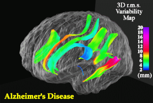
Results: 3D maps of regional atrophy and disease-specific abnormalities were generated for new AD subjects. Regionally-selective fiber loss in AD (with 24.5% loss in the midsagittal area of the corpus callosum's posterior midbody; p < 0.025) matched increases in structural variability in corresponding temporo-parietal projection areas. Confidence limits on 3D cortical variation exhibited severe increases in AD from 2-4 mm at the callosum to a peak standard deviation of 19.6 mm at the posterior left Sylvian fissure. Normal Sylvian fissure asymmetries (right higher than left; p < 0.0005) were accentuated in AD (p < 0.0002). 3D variability maps suggested left greater than right temporo-parietal degeneration in AD. AD-related ventricular enlargement and normal asymmetries were also mapped for the first time in 3 dimensions.
Conclusion: Complex alterations in cortical structure, found in AD and other diseases, underscore the need for brain atlasing systems which invoke a population-based analysis of structural variation to actively detect subtle and diffuse alterations in dementia.
References. Thompson PM et al.: [1]. J. Neuroscience (1996) 16(13):4261-4274; [2] IEEE Trans. Medical Imaging (1996) 15(4):1-16; [3]. J. Comp. Assist. Tomography (1997) 21(4):1-16.
Paul Thompson
| RESUME| E-MAIL ME| PERSONAL HOMEPAGE| PROJECTS |
|---|